RADIOGRAPHY
SOLUTIONS
Urgent Care
In the urgent care market, flexibility and speed are critical. Rayence offers a full line of X-ray equipment to meet the need of this market. Ranging from the cost-effective XR-5L to the innovative flexible Flex-Rad system, we have a solution to meet any requirement.
If a new x-ray room is not needed but throughput improvement is still the goal, Rayence has both tethered as well as wireless DR systems that can be easily added to any existing x-ray room in a matter of hours. All of our DR systems feature a full complement of DICOM protocols as well as software for in exam room viewing as well as burning of CDs.

Menu
DR Flat Panel Detectors
1417WCE - HR
1417WCC
1717SCV
Universal Radiography Systems
GOXR
Software
Xmaru Pro

1417WCE-HR
14x17 High Resolution Wireless Flat Panel Detector


Specifications
Sensor Type
Scintillator Type
Pixel Pitch (μm)
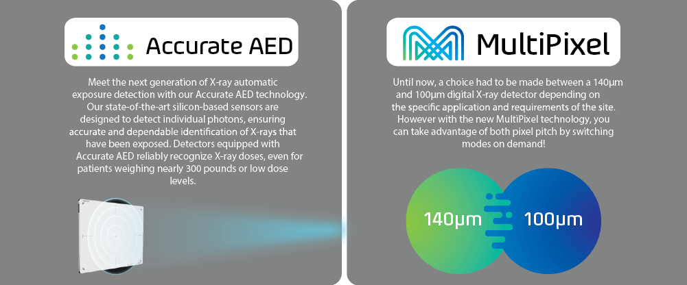
Accurate AED
MultiPixel
Glass Free
Battery Operating Time (hrs)
Weight (w/out battery)
Data Transfer
Energy Range (kVp)
A/D Conversion (bits)
Total Pixel Matrix (Pixels)
Dimension (in)
a-si TFT
Direct Deposit CsI
100
Yes
Yes
-
Max 16
5.2 lbs
GigE
802.11a/g/n/ac
40 - 150
16
3534 x 4302
15.1 x 18.1 x 0.6
* Specifications subject to change without prior notice *

1417WCC
14x17 Wireless DR Flat Panel Detector

PROVIDING PATIENT THROUGHPUT IN YOUR HOSPITAL AND BEYOND
The ergonomically designed C-Series Cesium Iodide wireless detectors are designed to offer new levels of handling, functionality and exceptional diagnostic image quality in the X-ray room and beyond.
The compact and lightweight Xmaru 1417 Wireless Digital Flat Panel Detectors are well designed to satisfy the daily diagnostic needs of the most demanding user.
DICOM File Management and Printing
Image Magnification
Image Stitching
CD recording with CD viewer
Measuring the Length and Angles of the Image
Adding Annotation Text, Graphics and Electronic Markers to an Image

Superb Image Quality
The detector's high Detector Quantum Efficiency (DQE) achieves superb image quality with low patient dose.
Durability
Supporting up to 660 lbs, the detector is manufactured with a shock, vibration, and scratch-resistant carbon fiber composition.
Water Resistant (IP 66)
The detector is water-resistant to most typical water spills in a hospital as well as in outdoor applicants.
Specifications
Active Area
Scintillator
Resolution
Effective Pixel Matrix
Pixel Pitch
A/D Conversion
Preview Time
Operation Environment
Energy Range
Data Output
Weight
Dimensions
16.8 x 13.8 inch
Cesium
3.5 lp/mm
2500 x 3052
140 ㎛
14/16 bit
≤ 5 Sec.
Temperature. +5 ~ +40°F
Humidity. 30 ~ 75%
Pressure. 70 ~ 106 kPa
40~150 kVp
Ethernet 1 Gbps Ethernet
6.6 lbs / 3 kg
18.1 x 15.1 x 0.6 in
* Specifications subject to change without prior notice *

1717SCV
17x17 Tethered Flat Panel Detector

UNIVERSAL AND AFFORDABLE SOLUTION WITH LARGE COVERAGE
The 1717SCV is designed to provide a larger imaging area while eliminating the need to rotate the detector to capture even the widest chest or abdomen.
The detector possesses the thickness of traditional ISO 4090 film cassettes, which makes retrofitting them into standard cassette trays easy. The use of 17x17 inch detector provides more flexibility when positioning anatomy and eliminates the need to be rotated for certain.
The 1717 S-Series is constructed with Rayence's auto-trigger signal sensing technology, removing the need for generator integration. Efficiency is improved by streamlining workflow through the elimination of the additional steps required when using film or CR.
Together with an image preview time of less than 5 seconds, patient throughput and overall productivity is increased, and the wait time for patients is decreased, providing an economical and ideal solution for any X-ray room.
No need an electrical connection
17x17 inch Active Area
Full field of view (43cm x 43cm)
Lightweight and Thin Dimensions
Reduced image preview time to 1.5 sec
140㎛ Pixel Pitch
Specifications
Detection Area
Sensor Type
Scintillator
Pixel Matrix
Pixel Pitch
A/D Conversion
Resolution
Preview Time
Energy Range
Data Output
Weight
Dimensions
17 x 17 inch
Amorphous Silicon with TFT
Cesium
3072 x 3072
140㎛
14/16 bit
Max. 3.5lp/mm
≤ 2Sec.
40~150kVp
Ethernet 1Gbps
9.25lbs / 4.2kg
18.1 x 18.1 x 0.6in
* Specifications subject to change without prior notice *

Digital Radiography Table


Versatile Full Featured Heavy Duty Radiographic Room
The GOXR is a full-featured radiographic room that comes standard with the Rayence’s 1417WCC or upgraded to premium with the NEW low dose GreenON DR system which is fully integrated with the generator controls and all DR and generator functions can be controlled from the large touch screen workstation display with the Xmaru Pro software.
Fully Integrated
Compact Design
Heavy Duty 6 Way Elevating Table
Recessed Foot-Pedals
Wall Stand Lowers to Below 10”
Cross Table Capability
The Elevating 6 Way Heavy Duty, all-steel table features a quiet
lift and a capacity of 650 lbs
There is also an option to order the system with an 84”x30.5”
tabletop and 4 way lock release
The 4 recessed foot pedal design simplifies
patient positioning eases accessibility, and
locking of tabletop movement. The collision
protection electronics and fail-safe locks ensure
safe operation
The ergonomic tube stand
handles are designed for
ease of positioning. Cross
table exams are easily
performed using the +/-
180 degree rotational and
+/- 5” transverse
positioning capability
The wall stand has quick-grip
release hand control for easy
positioning of the wall receptor
and side-mounted handgrip
bars stabilize the patient during
PA exams. (Optional overhead
patient handgrips help stabilize
patients for lateral views)
The long vertical travel of both
the wall stand and tube arm
facilitate a full range of exams
including weight-bearing
studies and the ergonomically
designed handle allows for easy
positioning of the wall stand
receptor
Specifications
DETECTOR
Model
TUBE STAND
Style
Rail length
Transverse travel
Long Travel of Focal Spot
Display
Wall to focal spot at detent position
Tube Rotation
WALL STAND
Model
Vertical Travel Range
Left or Right Hand Load Bucky
TUBE
Model
Tube Focal Spot
COLLIMATOR
Model
Style
Features
GENERATOR
Generator Power Output
mA range
Power
mAs range
TABLE
Model
Movement
Vertical travel
Transverse/Long Travel
Dimensions
Grid
OPTION
Option
Table travel
Transverse travel
Longitudinal travel
Weight Capacity
ROOM
Min. Required
1417WCC / GreenON 1417(Premium)
floor mounted
10 ft rail
9.75" travel
96"
angulation dial, handgrips, electric locks
Vertical Travel Range 14.56" - 72.44"
43.0"platform, 44.13" trunnion
180 degrees, 90 degree detents
J1000
15" min, 70" max
yes
E411
.6-1.5 focal, 200 KHU
5658
LED , 150kVp w/ swivel mount
laser bucky light and tape measure
421/3e, 42kW high freq
25-500mA
205/250-1 phase 208/408-3 phase
0.1-600mAs
S222
6 way elevating table
23-34"
10" transverse, 30" longitudinal
84x30", 74" table top option
103L, 10:1, 34-44"FD
downgrade to S223 4 way table
84x30", 51" long enclosed welded pedestal
plus or minus 5"
30"
400 lbs
12'6" x 11' x 9
* Specifications subject to change without prior notice *

XMARU PRO
Acquisition Software

SMART CHIROPRACTIC DIGITAL IMAGING SOLUTIONS
Xmaru PRO is the most flexible diagnostic acquisition software available for today’s healthcare providers. PRO uses the industry’s best image algorithm parameters for the highest quality diagnostic images while optimizing workflow with an easy to navigate GUI. Built on a 64 Bit platform takes advantage of improving hardware technology, speed, and improved image quality on larger displays.
HDR software improves observation
of Bones and Microstructures
GridON software reduces radiation scatter
Harmonic Automatic Stitching
Dual Exam Review-Images from two
independent studies can be shown side by
side for comparison

Intuitive GUI
Intuitive and Direct Graphic User
Interface with X-ray Detector and Generator
Optimized Exposure Conditions and Image Review
Reducing Unnecessary Rework and Test
Harmonic Stitching
(optional)
Image stitching is achieved by selecting one of three methods: Full-Auto, Semi-Auto or Manual. To eliminate the exposure borders of each image due to varying densities, Rayence’s advanced gradation process is automatically applied. Together with Rayence’s optional automatic stitching software, up to three views can be automatically stitched at a touch of a button, making stitching examinations easier than ever to attain.
System Requirement
Monitor Resolution
CPU
Memory
Operation System
1,280 x 768 (Minimum) 1,680 x 1,050 1,920 x 1,080 (Optimized)
Intel Core™ i5 or more
Intel GMA 950 or more Intel GMA X3500 or more Nvidia Geforce FX5200 or more 512MB or more (No Shared Memory)
Windows 7 (32bit/64bit) Professional or Home Edition Window 8 (32bit/64bit) Professional or Enterprise
* Specifications subject to change without prior notice *